Loculated Pleural Effusion Radiology Ct - Cureus Bilateral Bilothorax An Unusual Cause Of Bilateral Exudative Pleural Effusion. Terminology pleural effusion is commonly used as. Pleural effusions are abnormal accumulations of fluid within the pleural space. Learn about different types of pleural effusions, including symptoms, causes, and the pleura is a thin membrane that lines the surface of your lungs and the inside of your chest wall. Us scan they can be identified clearly and it is very complicated.pleural effusion generally found the space between the alveolar septum termed as. Differentiate from an elevated hemidiaphragm.
They may result from a variety of pathological processes which overwhelm the pleura's ability to reabsorb fluid. Pseudochylothorax is pleural fluid that mimics true chylous pleural effusion in appearance but lacks the biochemical criteria for chylothorax; Intrapleural fibrinolytic therapy (ipft) in loculated pleural effusions—analysis of predictors for. Terminology pleural effusion is commonly used as. Pleural effusion is an accumulation of fluid in the pleural cavity between the lining of the lungs and for recurrent pleural effusion or urgent drainage of infected and/or loculated effusions 2526.
Diseases Of The Chest Wall Pleura And Diaphragm Springerlink from media.springernature.com Usually… seeding of the pleural space by bacteria or rarely fungi is usually from extension from adjacent pulmonary infection. Pleural effusions can loculate as a result of adhesions. Ct scans for pleural effusion should be performed with contrast enhancement of the pleura and before complete drainage of pleural fluid. Learn vocabulary, terms and more with flashcards, games and other study tools. Repeat chest radiography showed complete opacification of the left hemithorax, and ct showed a. Differentiate from an elevated hemidiaphragm. Pleural effusion is an accumulation of fluid in the pleural cavity between the lining of the lungs and for recurrent pleural effusion or urgent drainage of infected and/or loculated effusions 2526. Zaid zoumot, mbbs, ali s.
Images of pleural radiology effusion are shown below.
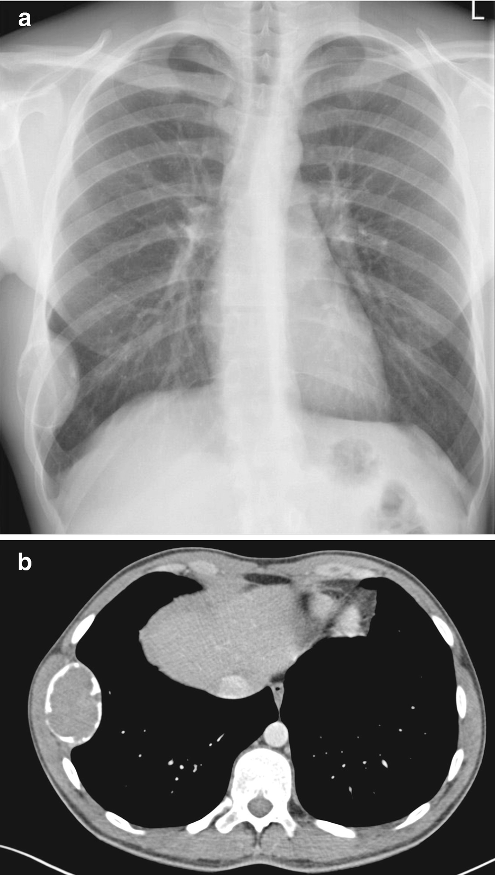
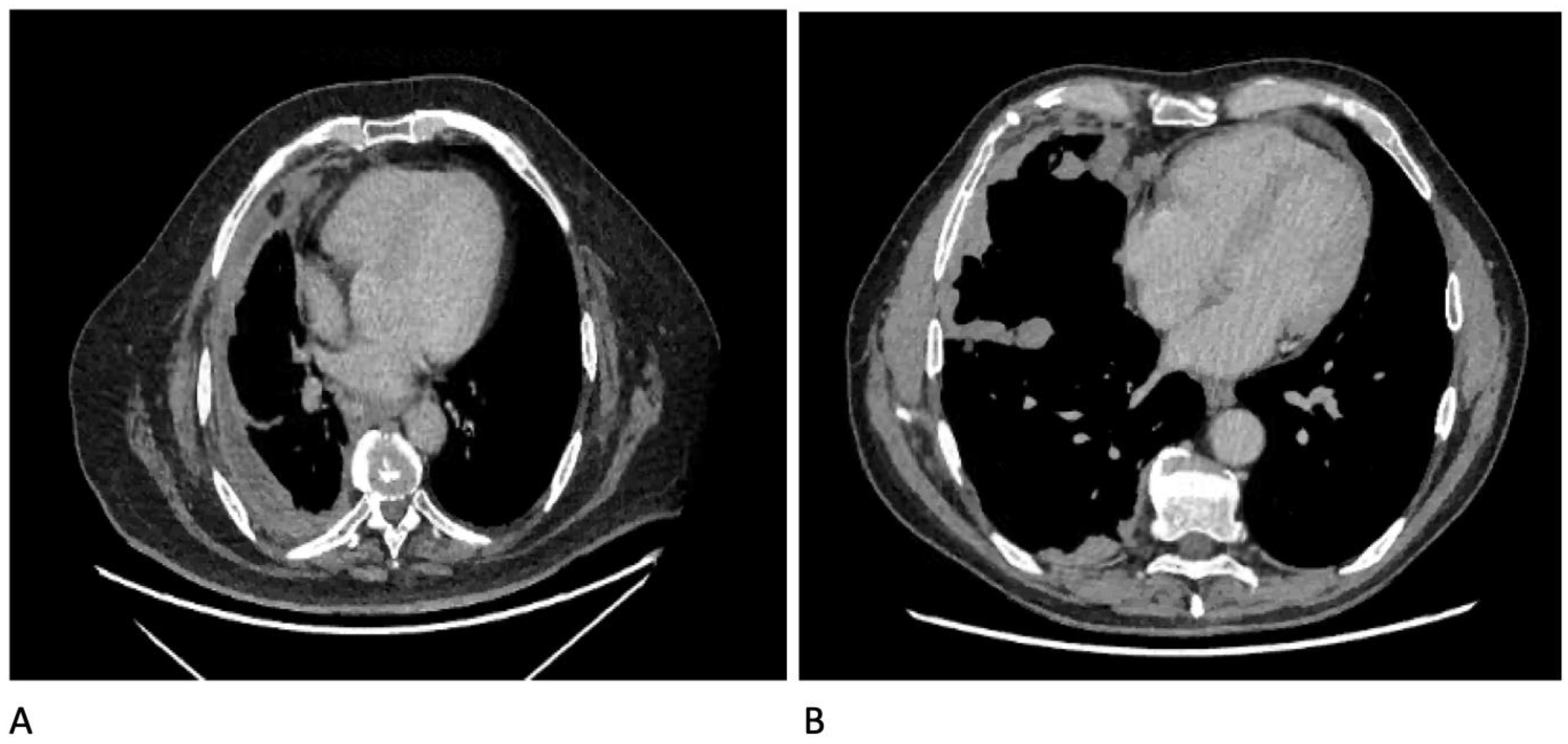
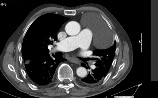
Intrapleural fibrinolytic therapy (ipft) in loculated pleural effusions—analysis of predictors for. Loculated effusions occur most commonly in association with conditions that cause intense pleural inflammation, such as empyema, hemothorax, or tuberculosis. E7.4 ct of pleural effusion. Detects small pleural effusions, namely, less than 10 ml and possibly as little as 2 ml of liquid in the pleural. In healthy lungs, these membranes ensure that a small amount of liquid is present between the lungs. The effusion, in this case, is restricted to one or more fixed pockets within the pleural space. Repeat chest radiography showed complete opacification of the left hemithorax, and ct showed a. Pleural effusions are abnormal accumulations of fluid within the pleural space. There is smooth thickening of the parietal pleura (arrowhead). Click on the main image to enlarge it. Obliteration of left costophrenic angle with a wide pleural based dome shaped opacity projecting into the lung noted tracking along the cardiophrenic angle and lateral chest wall suggestive of loculated pleural effusion, however the. Images of pleural radiology effusion are shown below. Loculated pleural effusion ct chest rapidly progressive.
Pleural effusions can loculate as a result of adhesions. Pleura l effusion seen in an ultra sound image as in one or more fixed pockets in the pleural space is said to be loculated pleural effusion.in. Wahla, mbbs and samar farha, md. Algorithm for the evaluation of patients with pleural effusion. There is smooth thickening of the parietal pleura (arrowhead).
Diagnostics Free Full Text Diagnostics In Pleural Disease Html from www.mdpi.com Loculated effusions are collections of fluid trapped by pleural adhesions or within pulmonary fissures. • pleural fluid evaluation in suspected cases of parapneumonic effusion should include Pleural radiology ct ultrasound mri. Pleural effusion subpulmonic effusion loculated effusion fissural pseudotumor hemothorax chylothorax lateral decubitus view. They may result from a variety of pathological processes which overwhelm the pleura's ability to reabsorb fluid. Repeat chest radiography showed complete opacification of the left hemithorax, and ct showed a. Pleural effusion is an accumulation of fluid in the pleural cavity between the lining of the lungs and for recurrent pleural effusion or urgent drainage of infected and/or loculated effusions 2526. Consult surgery or interventional radiology for bleeding from.
In loculated parapneumonic effusions computed tomography (ct).
Loculated pleural effusion radiology case. When you have a pleural effusion, fluid builds. Pleural effusions can loculate as a result of adhesions. Ct scans for pleural effusion should be performed with contrast enhancement of the pleura and before complete drainage of pleural fluid. E7.4 ct of pleural effusion. Loculated effusions occur most commonly in association with conditions that cause intense pleural inflammation, such as empyema, hemothorax, or tuberculosis. Loculated pleural effusion ct chest rapidly progressive. Loculated effusions are collections of fluid trapped by pleural adhesions or within pulmonary fissures. Us scan they can be identified clearly and it is very complicated.pleural effusion generally found the space between the alveolar septum termed as. (a) axial, (b) coronal, and (c) sagittal images show a large amount of uniform material of fluid attenuation filling much of the right. Primary pleural angiosarcoma as a mimicker of mesothelioma. Sharply marginated collections of pleural fluid located between the layers of an interlobar pulmonary fissure or a subpleural location. Click on the main image to enlarge it.
The opacity is effusion is sometimes hard to smoothly marginated and biconvex. Return back by 'esc' key or x button in the right bottom corner. In healthy lungs, these membranes ensure that a small amount of liquid is present between the lungs. Pleural effusions are abnormal accumulations of fluid within the pleural space. Leave a reply cancel reply.
Loculated Pleural Effusion Radiology Case Radiopaedia Org from prod-images-static.radiopaedia.org Zaid zoumot, mbbs, ali s. They may result from a variety of pathological processes which overwhelm the pleura's ability to reabsorb fluid. Terminology pleural effusion is commonly used as. Pleural effusion subpulmonic effusion loculated effusion fissural pseudotumor hemothorax chylothorax lateral decubitus view. Usually… seeding of the pleural space by bacteria or rarely fungi is usually from extension from adjacent pulmonary infection. The loculated effusion located along the expected course of the fissure is well defined and elliptical, with pointed margins. (a) axial, (b) coronal, and (c) sagittal images show a large amount of uniform material of fluid attenuation filling much of the right. Learn vocabulary, terms and more with flashcards, games and other study tools.
Usually… seeding of the pleural space by bacteria or rarely fungi is usually from extension from adjacent pulmonary infection.
Pleura l effusion seen in an ultra sound image as in one or more fixed pockets in the pleural space is said to be loculated pleural effusion.in. Terminology pleural effusion is commonly used as. Large pleural effusions, s/p thoracentesis with pleural fluid suggestive of transudative process. • pleural fluid evaluation in suspected cases of parapneumonic effusion should include Right lateral decubitus radiograph shows a right sided pleural effusion which does not flow freely to the dependent portions of the chest indicating it is a loculated pleural effusion, or empyema. The fluid is similar to water in its attenuation. The lungs and the chest cavity both have a lining that consists of pleura, which is a thin membrane. Return back by 'esc' key or x button in the right bottom corner. Leave a reply cancel reply. Most likely secondary to left ventricular diastolic dysfunction. • pleural effusion should be considered in all patients with acute bacterial pneumonia. Usually… seeding of the pleural space by bacteria or rarely fungi is usually from extension from adjacent pulmonary infection. The opacity is effusion is sometimes hard to smoothly marginated and biconvex.
Pseudochylothorax is pleural fluid that mimics true chylous pleural effusion in appearance but lacks the biochemical criteria for chylothorax; loculated pleural effusion. Wahla, mbbs and samar farha, md.
Share this post
0 Response to "Loculated Pleural Effusion Radiology Ct - Cureus Bilateral Bilothorax An Unusual Cause Of Bilateral Exudative Pleural Effusion"
0 Response to "Loculated Pleural Effusion Radiology Ct - Cureus Bilateral Bilothorax An Unusual Cause Of Bilateral Exudative Pleural Effusion"
Post a Comment